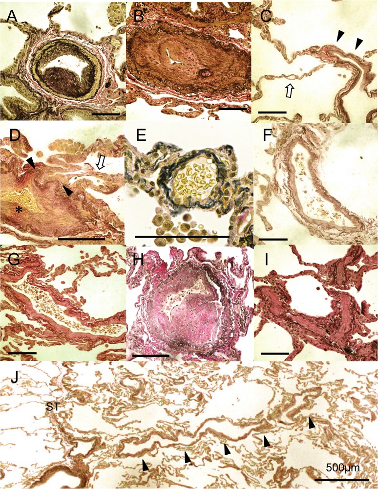
Fig 1. Representative photographs of the pulmonary arterioles, the pre- and post capillary vessels and the pulmonary venules.
(A) Fibrous intimal thickening and mild medial thickening of the pulmonary muscular arteries (Case 2). (B) Cellular intimal thickening of the pulmonary muscular arteries resulting in obstruction of the lumen (Case 4). (C) Remodeling of post capillary vessels (arrow heads) (Case 10). The structures of capillaries did not show hemangiomatous appearance (arrow). (D) A remodeling lesion in the postcapillary vessels (arrow heads). The capillaries collapsed due to the obstruction of the proximal vessels (skeleton arrow) (Case 7) (*: Initimal fibrous thickening of a pulmonary vein is shown). (E) A venule next to the interlobular septa without intimal thickening (PV score 0) (Case 7). (F) A pulmonary vein with slight intimal thickening next to the interlobular septa (PV score 1) (Case 10). (G) A pulmonary venule with fibrotic intimal thickening (PV score 2) (Case 9). (H) A muscularized pulmonary vein with marked intimal thickening and moderate medial alterations. It is similar to the structure of pulmonary muscular arteries (Score 3) (Case 1). (I) Pulmonary venules with severe intimal thickening resulting in luminal obstruction similar to the pathology of pulmonary veno-occlusive disease (PVOD) (PV score 4) (Case 4). (J) The remodeling seems to extend from the post capillary vessels to the interlobular septum (Case 10). (All slides were stained with Elastica van Gieson stain. Scale bars shows 100 μm unless otherwise stated)

