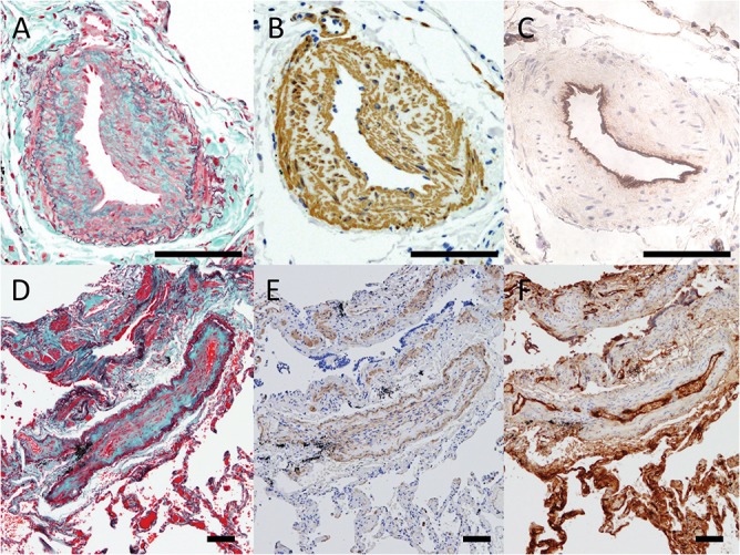
Fig 2. Modified Masson-Goldner stain and immunohistochemistry of pulmonary arteriopathy and venopathy.

A to C show pulmonary muscular arteries from Case 17 with intimal fibrotic changes and moderate medial thickening. (A) Modified Masson-Goldner stain (Elastica Masson stain) shows the complicated arrangement of collagen (green), elastin (dark-purple) and spindle cells (red) within the intima. (B) Numerous α-smooth muscle actin (SMA) positive cells are recognized in the thickened intima and media in the artery. (C) Factor VIII is negative within the intima and media. Only endothelial cells are positive for factor VIII. D to F show pulmonary venules from Case 10 with intimal alterations. (D) Modified Masson-Goldner stain show that the neointima of venopathy is rich in collagen (green). (E) α-SMA-positive cells can be seen in the media and thin intima; however, the number of the cells is less than that seen in pulmonary arteriopathy. (F) Factor VIII is only positive in the endothelial cells. (Scale bars show 100 μm)
