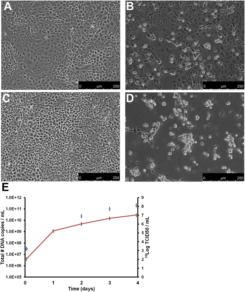
Fig 2. Replication of SDDV on SK21 cells.
SK21 cells were inoculated with an MOI of 0.01 TCID50/cell SDDV (passage 3). (A/C) Negative control confluent monolayer of SK21 cells on day 3 (A) and 4 (C); (B/D) SDDV-infected SK21 cells on day 3 (B) and 4 (D) after inoculation; (E) SDDV genome copy number/mL (red line and left y-axis) and infectious titer (blue diamonds and right y-axis) of the infected SK21 cells in time. Error bars represent the standard deviation.

