
Fig 3. Electron microscopy of SDDV.
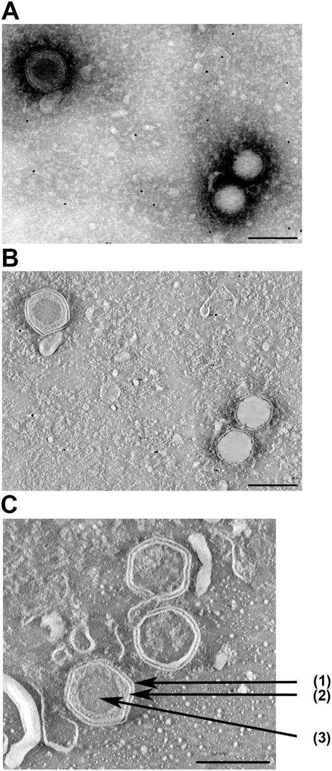
Hexagon particles with (upper left) or without (lower right) outer membrane as visualized by by negative staining (A) followed by tomography (B). In (C) the outer membrane (1), capsid (2) and inner core (3) of a different enveloped particle are indicated by arrows. In (B) and (C) (ortho)slices through virus particles are shown.