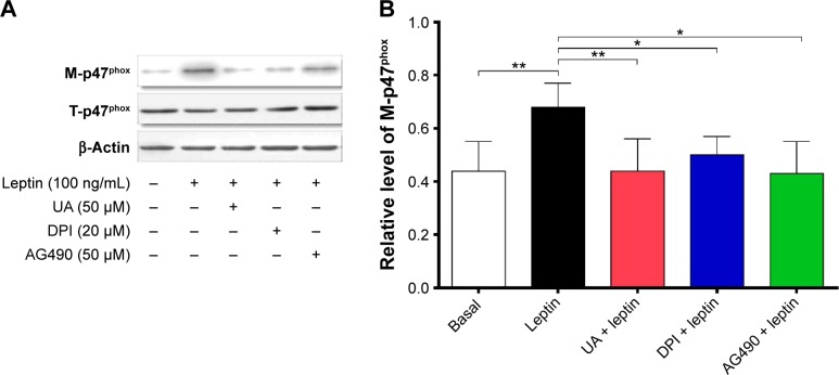
Figure 7.
UA suppressed leptin-induced translocation of the p47phox from cytoplasm to cell membrane in HSC-T6 cells.
Notes: Cells were pretreated with UA (50 µM), DPI (20 µM), or AG490 (50 µM) for 30 minutes and then were stimulated by leptin (100 ng/mL) for another 30 minutes. The membrane bound and total level of p47phox were examined by Western blotting assay. (A) Representative blots of membrane (M-) p47phox and total (T-) p47phox in HSC-T6 cells. (B) Bar graph showing the relative level of M-p47phox in HSC-T6 cells. Data are expressed as mean ± SD from six independent experiments. β-Actin acts as an internal control. *P<0.05; **P<0.01.
Abbreviations: UA, ursolic acid; HSC, hepatic stellate cell; DPI, diphenyleneiodonium; SD, standard deviation.

