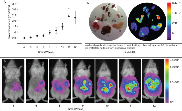
Fig 2. Tumour growth monitored by Bioluminescence Imaging (BLI).
Tumour growth was monitored weekly by in vivo BLI and an increase in the net bioluminescence versus time was observed (A, B). Organs were also examined by BLI post-mortem to visualize metastatic spread (C). Strong BLI signals were detected at site of injection (left uterine horn; luh), right ovary (o), connective tissue surrounding the uterine horn (ct), pancreas (p) and metastatic node (mn). Spot signals were detected in the liver (l), spleen (s), kidneys (k), heart (h) and lung (lu). No signal was detected in adrenal gland (a).

