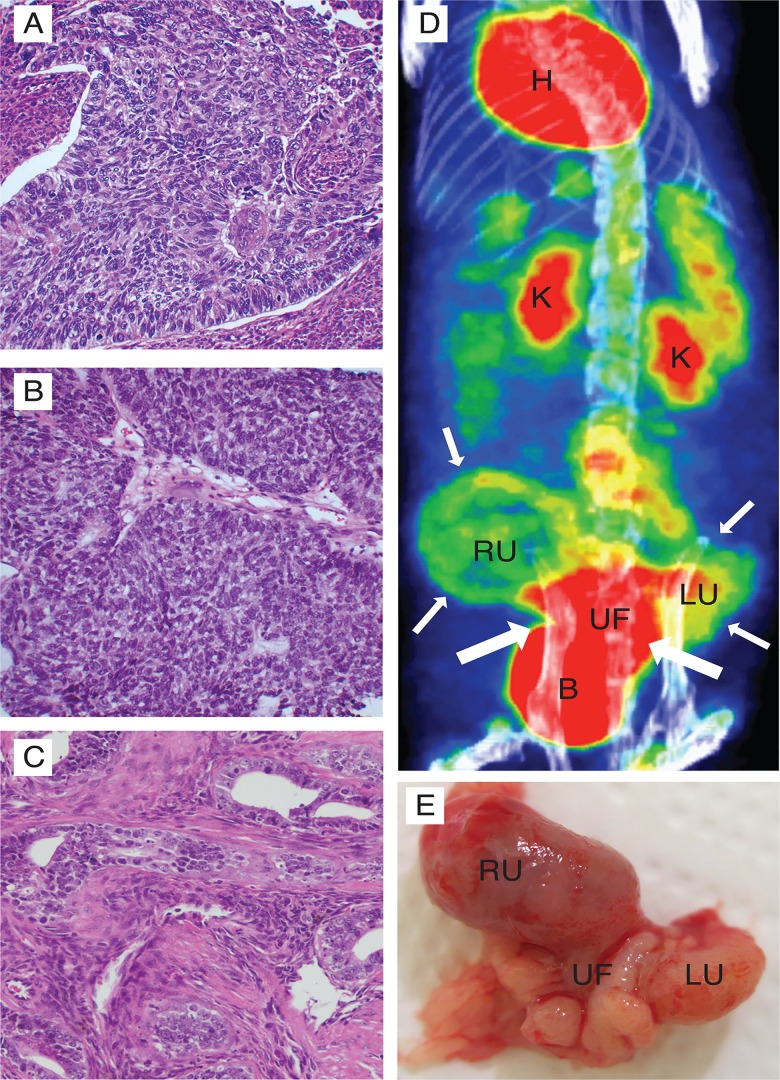
Fig 6. 18F-FDG PET detects tumour in an orthotopic endometrial cancer PDX model.
Mice were implanted in the uterus with cancer cells from a patient biopsy and rederived for two generations to develop a PDX model. Histological examination revealed that both the F1 (B) and the F2 (C) mice developed tumours closely resembling the parental tumour, defined as an endometrioid grade 3 endometrial cancer (A). 18F-FDG PET was successfully used to detect tumour growth in both generation F1 (not shown) and generation F2 (D). Highly 18F-FDG-avid tumour tissue (large arrows) in the uterine fundus (UF) and both the left and the right uterine horn (LU, RU respectively) depicted at PET-CT (D). Macroscopic examination revealed a large tumour infiltrating both the uterine horns as well as the bladder (E) and corresponded well with the tissue detected as tumour by 18F-FDG PET. B, bladder; H, heart; K, kidney; LU, left uterine horn; RU, right uterine horn; UF, uterine fundus.

