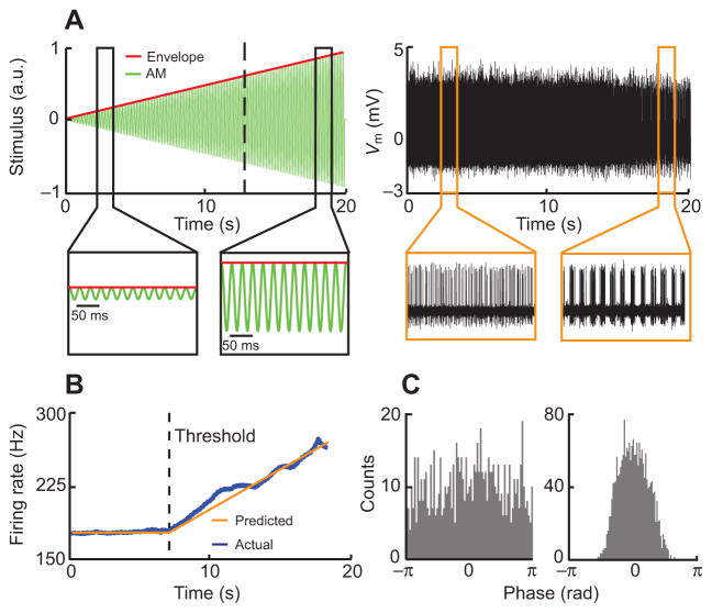
Fig. 6.
Peripheral electrosensory neurons respond to envelopes. (A) Left: example stimulus in which the envelope (red) increases linearly as a function of time with a 20 Hz sinusoidal AM (green). a.u., arbitrary units. Right: example recording from an electroreceptor in response to this stimulus. It can be seen that the electroreceptor phase locks to high but not to low envelopes. Vm, membrane potential. (B) Peri-stimulus time histogram (PSTH) from the same electroreceptor. It can be seen that the firing rate is constant for low envelope values and increases linearly when the envelope is greater than a threshold value (dashed line). (C) Phase histograms when the envelope is lower than threshold (left) and higher than threshold (right). Phase locking (i.e. the absence of firing at some carrier phases) is observed only when the envelope is higher than threshold.

