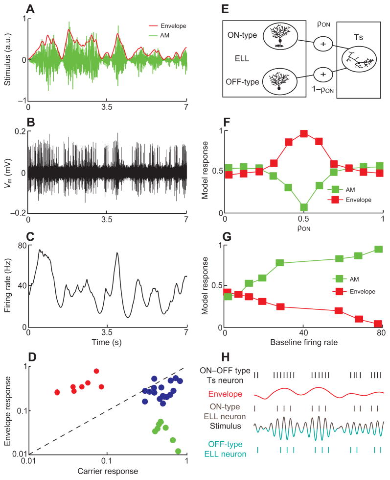
Fig. 8.
Midbrain electrosensory neurons respond to envelopes. (A) The stimulus consists of a noisy AM (green) with corresponding envelope (red).
(B) Response from an example Ts neuron to the stimulus shown in A. This neuron responds strongly to the envelope. (C) PSTH response from this same Ts neuron to the stimulus shown in A. (D) Envelope response as a function of AM response from Ts neurons. In contrast with ELL pyramidal cells, three distinct clusters are seen. Some Ts neurons respond selectively to either the envelope (red) or the AM (green) while some respond to both (blue). (E) Model in which a Ts neuron receives input from both ON- and OFF-type ELL pyramidal cells. The strength of the input from ON-cells is given by ρON while the strength of the input from OFF-cells is given by 1–ρON. Both inputs are thus balanced in strength when ρON=0.5. (F) Model results showing the AM (green) and envelope (red) responses as a function of ρON. (G) Model results showing the AM (green) and envelope (red) responses as a function of the baseline firing rate. (H) Proposed schematic diagram by which Ts neurons acquire their response selectivity to envelopes. ON-and OFF-type ELL pyramidal cells respond during the positive and negative phases of the AM, respectively (brown and green). However, both tend to respond more when the envelope is high. Summing these responses gives a response that is independent of AM but will still depend on the envelope (black).

