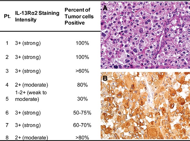
Figure 1.

IL-13Rα2 staining intensity and percent positive cells for each enrolled patient with metastatic ACC. (A) Representative hematoxylin and eosin (H&E) staining of patient tumor. (B) Representative IL-13Rα2 staining of patient tumor. IL, interleukin; ACC, adrenocortical carcinoma.
