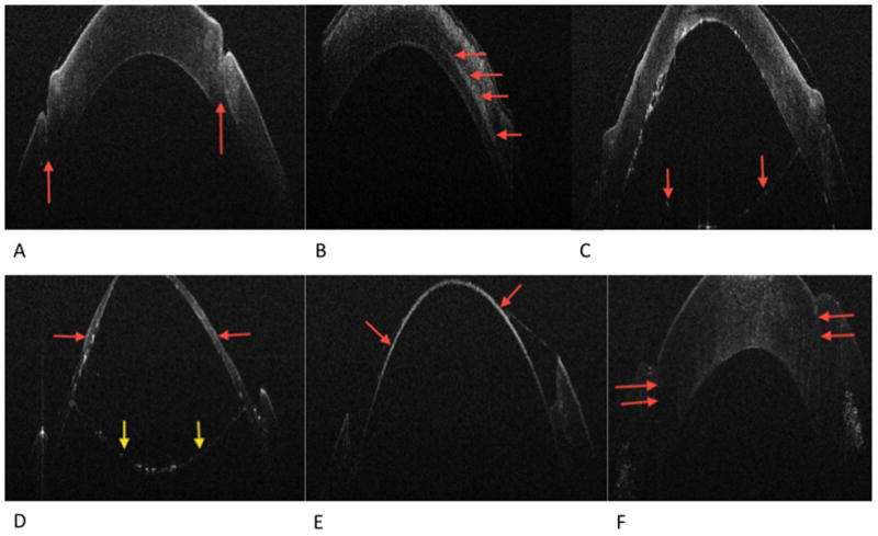
Figure 2.

Figure 2A. Post-host partial thickness trephination. The red arrows indicated trephination groove
Figure 2B. Post-cannula tunnel creation. The red arrows indicated cannula tunnel once the Fogla pin was removed
Figure 2C. Post-big-bubble. The red arrows indicated the Descemet’s membrane/endothelial complex separated by the overlying stroma by air
Figure 2D. Post-initial anterior stromal manual dissection. The red arrows indicated residual stroma following the initial anterior lamellar dissection. The yellow arrows indicate the Descemet’s membrane/endothelial complex
Figure 2E. Post final dissection, bared Descemet’s membrane indicated by the red arrows
Figure 2F. Post-graft suturing. The red arrows indicate the graft host stromal apposition. There is an apparent anterior wound gap, which actually represents the donor edge tucked under
