Fig 2.


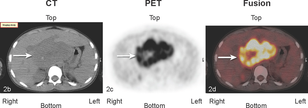
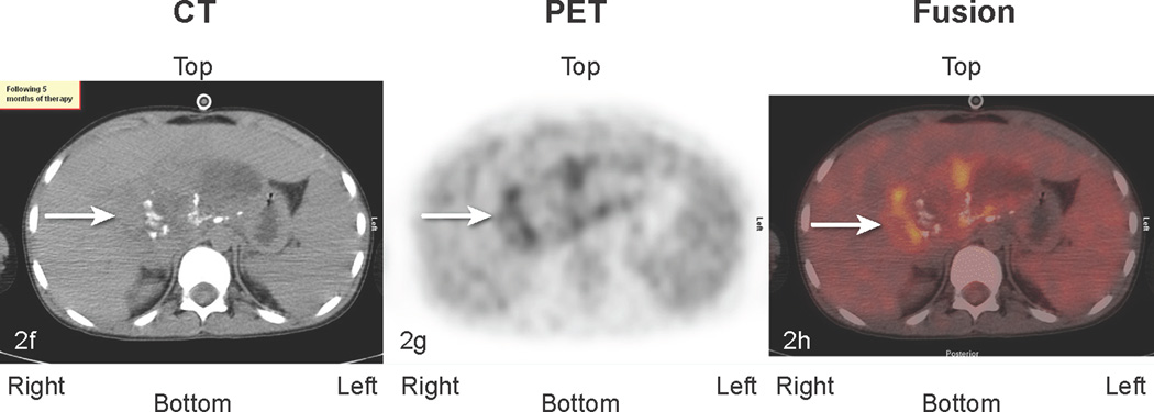
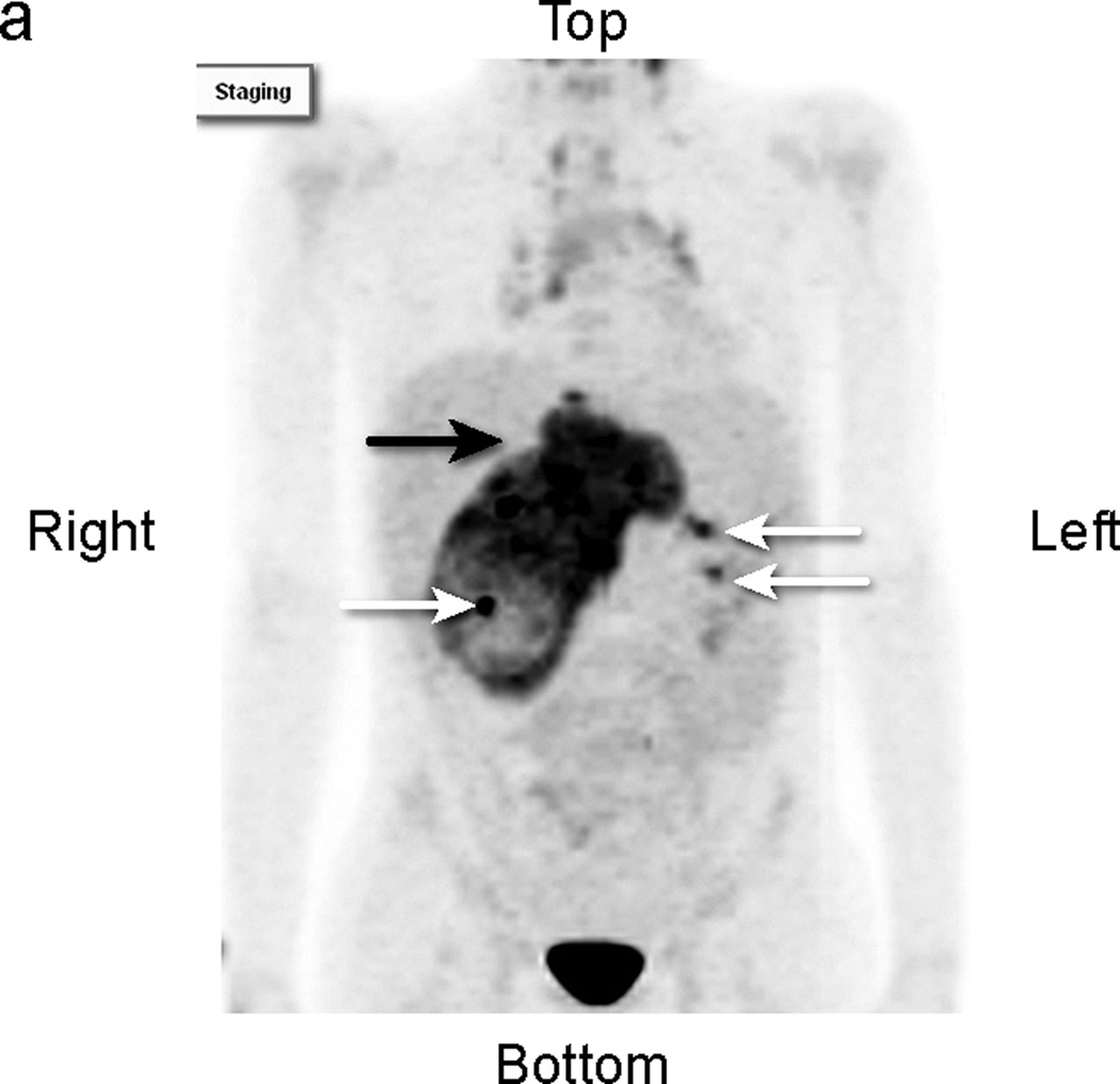
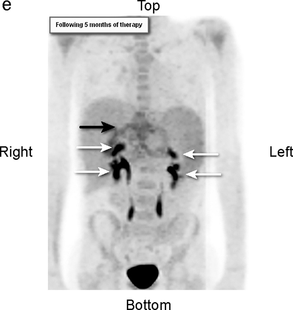
Patient # 3: 11-year-old boy with DSRCT and FDG-avid mass in his abdomen and thoracic lymph nodes. Anterior MIP image from FDG PET/CT scans at a) initial staging shows a very large accumulation of intense activity (black arrow) in the right side of the abdomen and scattered foci of uptake in the chest. White arrows point to renal activity. b,c,d) transverse CT, PET, and fusion images of the mid abdomen show a large area of markedly elevated uptake (white arrows). e) Anterior MIP image shows considerable reduction in mid abdominal activity (black arrow). White arrows show renal accumulation. f,g,h). Transverse CT, PET, and fusion images of the mid abdomen show residual uptake (white arrows) within the central tumor mass, partly calficied. The extent and intensity of uptake have declined considerably.


