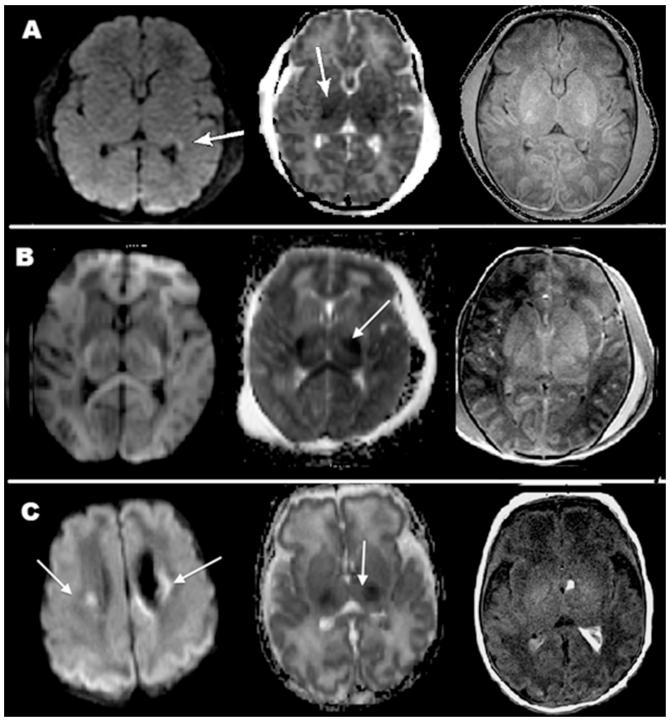
Figure 1.
Magnetic resonance imaging (MRI) images in three patients showing similar findings despite different clinical outcomes. (A) Patient 1: Restricted diffusion in parietal/occipital/temporal white matter and thalami on day of life (DOL) 4. The patient had normal neurological outcome at 4 months; (B) Patient 12: Restricted diffusion in bilateral thalami on DOL 5. The patient is deceased; (C) Patient 11: Restricted diffusion in bilateral thalami, corona radiata and parietal/occipital cortex. At 36 months the patient had mild developmental and speech delay.

