
Fig. 2.
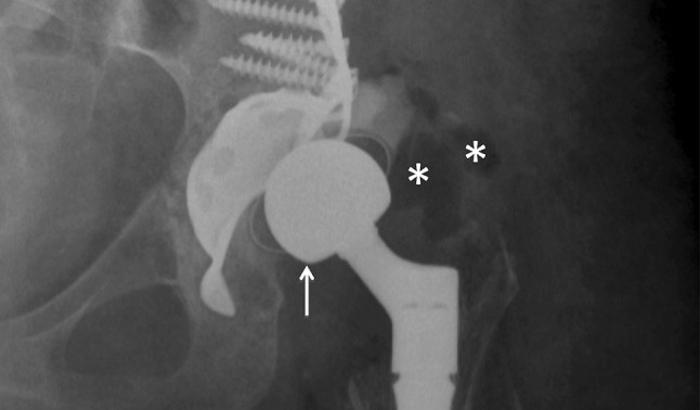
X-ray of a patient with hip dislocation and suspected infection. Antero-posterior projection shows cranial dislocation of implant femoral head (white arrow) and peri-prosthetic soft tissues swelling. Asterisks show air within peri-prosthetic soft tissues