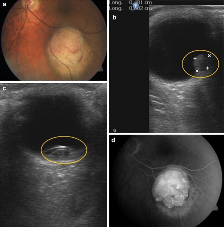
Fig. 7.
a Ophthalmoscopic image of choroidal amelanotic melanoma involving the lower temporal quadrant of the eye. b Corresponding ultrasonography image of choroidal amelanotic melanoma. c Corresponding ultrasonography image of choroidal amelanotic melanoma (different projection). d Corresponding angiography with fluorescein

