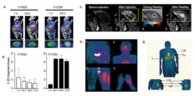
Figure 1. Image-guided drug delivery.
A-B: At the preclinical level, image-guided drug delivery can be used for several different purposes, including for the analysis of active vs. passive tumor targeting. Panels A and B show that at early time point after i.v. administration, RGD-modified tumor vasculature targeted polymeric drug carriers accumulate more efficiently within tumors than peptide-free passively targeted polymers. They also show, however, that over time, the latter are more efficient in achieving high tumor concentrations. C: MR imaging of gadolinium release from temperature-sensitive liposomes before and after heating, exemplifying that image-guided drug delivery can be used to tailor triggered drug release. D-E: Accumulation of radiolabeled liposomes in tumors in patients, showing that sarcomas tend to accumulate nanomedicine formulations relatively well (D), whereas breast carcinomas present with a relatively low degree of EPR-mediated tumor accumulation (E). Tu: tumor, LN: lymph node, L: liver, Spl: spleen. Images adapted, with permission, from [8,10,11,12].

