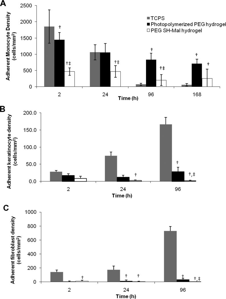
FIGURE 9.
Adherent cell density on TCPS, photopolymerized PEG hydrogel and PEG SH-Mal hydrogel. Three cell types were included, primary human monocyte (A), keratinocyte (B), and fibroblast (C). Cells were observed at 10× magnification. Five images per sample were taken at random fields of view. †, p < 0.05 vs. the cell adhesion density on TCPS at the same time point; ‡, p < 0.05 vs. the cell adhesion density on photopolymerized PEG hydrogel at the same time point. All data presented as average ± standard deviation (n=3).

