
Figure 1.
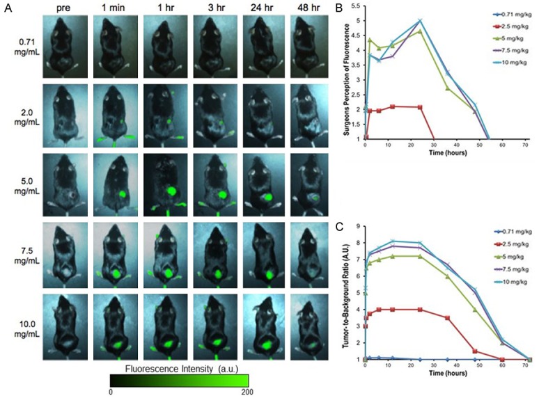
A. Subcutaneous tumors are imaged in vivo through the skin on the flank of mice. Tumor fluorescence with variation to ICG dosage and time dictate how it is optimally viewed. Increasing dosage leads to brighter fluorescence. With respect to time, the optimal fluorescence increased and peaked at 24 hours post injection, then steadily declined over the course of the next two days. B. Surgeons could not visualize the fluorescence from lower doses. At 5 mg/kg to 10 mg/kg, the surgeons did not subjectively perceive any difference in fluorescence. C. Tumor fluorescence was measured by region-of-interest software.