
Figure 6.
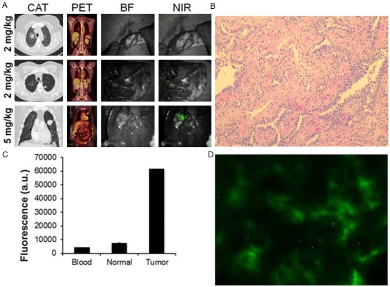
A. Representative images of three patients who underwent intraoperative imaging with ICG. Each patient underwent preoperative computed axial tomography (CAT) and positron emission tomography (PET) scanning. During the operation, each case was photo-documented by brightfield (BF) and NIR light. The first patient received 2 mg, the second patient received 2 mg/kg, and the third patient received 5 mg/kg. B. H&E and fluorescence microscopy confirmed the ICG was accumulating in the tumor tissue. C. Tumor, normal lung and blood fluorescence was measured by spectroscopy