Figure 4.

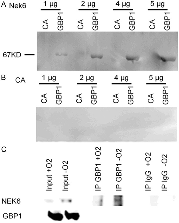
Representative far western blot showing the GBP-1: Nek6 interaction. In A GBP-1 was spotted onto the membrane. Carbonic anhydrase (CA) and recombinant Nek6 was incubated at 4 concentrations (from 1 to 5 µg). Interaction was revealed with an anti-Nek6 antibody. A clear band at the expected weight of GBP-1 (67kD) was seen at all the four concentrations with Nek6 but not with CA. In B the same experiment was repeated by spotting CA onto the membrane. No signal was detected with either Nek6 or CA at all concentrations. This experiment was repeated thrice with the same results. In C results of co-IP performed in A2780 cells in normoxic or hypoxic conditions (72 h). On the left input signal for both Nek6 and GBP-1; in the middle, pulldown of the GBP-1 protein and revelation with the anti-Nek6 identifies a signal which is higher in hypoxic conditions; on the right no signal is detected when the pulldown is performed with an anti-human-IgG. This experiment has been repeated thrice with similar results.
