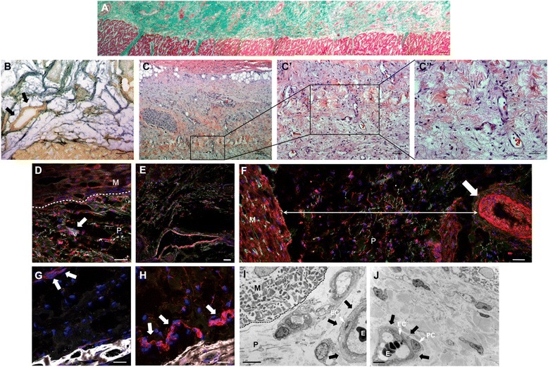
Fig. 3.

Neovascularization of pericardial-derived scaffolds after myocardial infarction. a Masson’s trichrome staining composition screening demonstrated the correct adhesion of the cardiac bioimplant with the adjacent myocardium. b and c-c′′ Gallego’s modified trichrome and hematoxylin/eosin staining exhibiting vessels in the cardiac matrix and zoomed images, respectively. Immunofluorescence analysis in the pericardial-derived scaffold after its implantation against isolectin B4 (green), smooth muscle actin (red), and elastin (white) indicates the presence of microvasculature (d), venules (e), and arterioles (f). Red positive immunostaining against CD31 (g) and von Willebrand factor (h) antibodies in pericardial-derived scaffold (arrows). Nuclei are counterstained with 4′,6-diamidino-2-phenylindole (DAPI) (blue) (i and j). Transmission electron microscopy images of the pericardial-derived scaffold show vascular structures (arrows) where endothelial cells (EC), pericytes (PC), and erythrocytes (E) are present. M myocardium, P pericardial-derived scaffold. Scale bars = 100 μm (a-c), 50 μm (c′ and d-h), 20 μm (c′′), and 5 μm (i and j)
