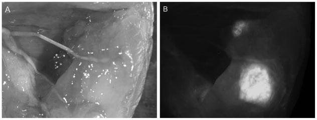
Figure 4. Fluorescence-guided resection of tumors.

By probing with a fluorescently-labeled antibody for a tumor marker, surgeons can immediately determine whether a tumor has been fully resected, and identify local metastases that may not be visible by eye. Here, a far-red fluorescent dye (Cy5)-labeled anti-PSCA diabody was used to guide resection of an intramuscular 22rv1-PSCA prostate cancer xenograft in a murine model. Cy5-anti-PSCA diabody was injected intravenously and surgery was performed 6 hours post-injection. (A) A white-light image of the tumor bed post-resection, with residual tumor purposely left unresected. (B) Fluorescence imaging shows residual tumor clearly. (Behesnilian et al., 2015)
