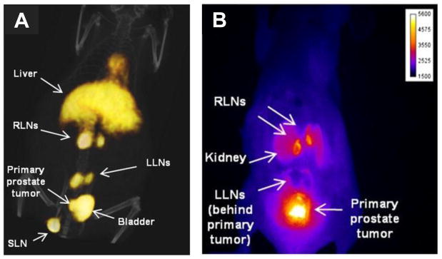
Figure 5. Dual-modality antibody imaging.

Non-invasive staging of lymph nodes (LN) followed by image-guided resection of only cancer-positive LNs offers more precision in identifying and removing metastases. Hall et al. used a dual-labeled anti-EpCAM mAb, conjugated with both IRDye 800CW and 64Cu-DOTA, to image prostate cancer LN metastases with PET/CT (A) and NIR fluorescence (B) imaging. PC3 cells were implanted in the prostate and imaging was performed 10–12 weeks later. Lumbar LNs (LLNs); renal LNs (RLNs); sciatic LN (SLN). This research was originally published in JNM. Hall MA et al. J Nucl Med. 2012;53:1427–37. © by the Society of Nuclear Medicine and Molecular Imaging, Inc.
