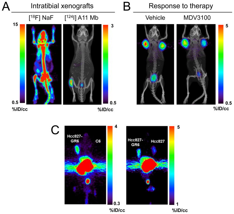
Figure 6. Applications of antibody imaging in oncology.
Imaging of specific biomarkers can be used to accurately locate lesions, aid in choosing a treatment, and monitor response to therapy. (A) PET imaging using the 124I-anti-PSCA minibody A11 was able to detect intratibial LAPC-9 prostate cancer xenografts with higher sensitivity and specificity than a 18F-Fluoride bone scan. (B) Response to anti-androgen treatment was monitored by visualizing reduced 124I-A11 activity in the tumors of enzalutamide-treated mice, reflecting a therapy-induced downregulation of PSCA expression. (C) The Hcc827-GR6 non-small cell lung cancer line developed resistance to gefitinib through upregulation of MET expression. PET imaging MET using a 89Zr-DFO-labeled MET-specific human minibody clearly visualized the overexpression of MET in the gefitinib-resistant tumors. (Knowles et al., 2014a; Li et al., 2014)

