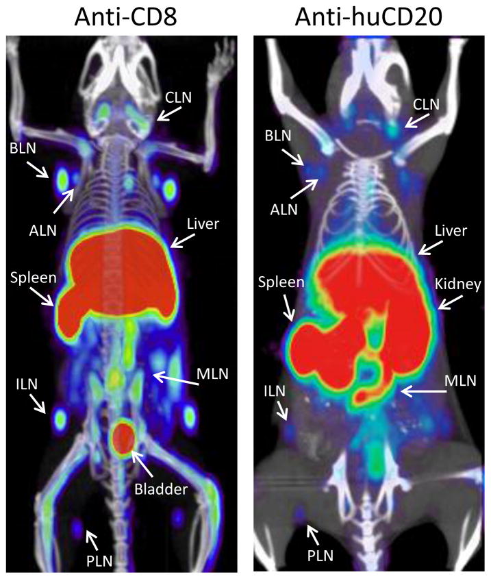
Figure 7. Imaging immune cell subsets.
Tracking CD8+ T cells could help detect and stage CD8+ lymphomas and aid in monitoring T cell immunotherapy. Here, imaging of CD8+ T cells with a 64Cu-NOTA-anti-CD8 minibody in wild type mice visualizes the spleen and lymph nodes (left panel). 89Zr-DFO-anti-huCD20 cys-minibody imaging of a transgenic mouse expressing huCD20 reveals B cells in the spleen and lymph nodes (right panel); this could also be applied to for the detection of B-cell lymphomas. (Tavaré et al., 2014a; Zettlitz et al., 2013)

