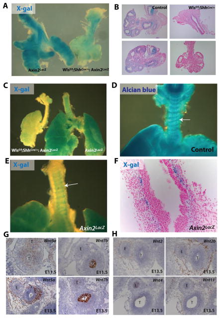
Figure 2. Decreased mesenchymal canonical Wnt signaling after deletion of epithelial Wls.
X-gal staining was performed on trachea-lung explants of E12.5 Axin2LacZ and Wlsf/f;ShhCre/+;Axin2LacZ mice. X-gal staining was primarily observed in mesenchyme of Axin2LacZ mice, while staining was nearly absent in tissue of Wlsf/f;ShhCre/+;Axin2LacZ mice (A,B). At E14.5, X-gal staining was detected in the tracheal mesenchyme of control embryos in a pattern similar to that of the cartilaginous rings. In contrast, no staining was detected in the trachea of Wlsf/f;ShhCre/+ embryos (C). E14.5 control tracheal explants were stained with Alcian blue to determine sites of chondrogenesis. The staining pattern is similar to the X-gal staining of Axin2LacZ mice (D). X-gal staining was restricted to the periphery of tracheal mesenchymal condensations (E, F). In situ hybridization was performed to determine expression pattern of several Wnt ligand mRNA. At E11.5 Wnt5a was detected in both epithelium and mesenchyme of developing trachea. At E13.5 Wnt5a RNA was enriched in the ventral mesenchyme of developing trachea, while Wnt7b was restricted to the epithelium of E11.5 and E13.5 trachea (G). Wnt2 and Wnt2b mRNA were detected in dorsal and lateral mesenchyme surrounding the trachea. Wnt4 mRNA was detected at low levels in the tracheal epithelium, while Wnt11 was present in tracheal mesenchyme of E13.5 embryos (H).

