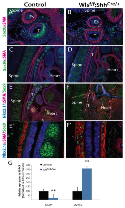
Figure 3. Loss of epithelial Wls-mediated signaling disrupts dorsal-ventral patterning of the trachea.

Longitudinal and cross sections of E13.5 (A,B) and E14.5 (C,D,E,F) embryos were stained with Sox9, aSMA and Nkx2.1 antibodies. DAPI was utilized to visualized cell nuclei. Sox9 was absent in the tracheal mesenchyme but expressed in vertebre cartilage of Wlsf/f;ShhCre/+ embryos. αSMA staining was expanded into the ventral region of the trachea of Wlsf/f;ShhCre/+ embryos (B,D,F). In developing trachea of Wlsf/f;ShhCre/+ embryos, muscle fibers run parallel to the longitudinal axis of the trachea in contrast to the transversal arrangements of the fibers observed in control trachea (E′, F′). Sox9 was decreased and Acta2 mRNA increased in tracheal explants of Wlsf/f;ShhCre/+ embryos at E14.5 (G). N=4, **p<0.01, T-trachea Es= esophagus.
