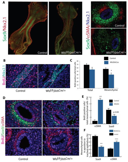
Figure 5. Epithelial Wnt ligands modulate mesenchymal cell proliferation.
Confocal images of E11.5 tracheal lung explants stained for Sox9 and Nkx2.1 are shown. Sox9 staining was nearly absent in the tracheal mesenchyme of Wlsf/f;ShhCre/+ mice. Transverse sections of tracheal tissue depict the extent of Sox9 expression and the lack of aSMA staining in Wlsf/f;ShhCre/+ at E11.5. Dams were injected at E11.5 with BrdU to determine cell proliferation. Longitudinal sections of E12.5 tracheas were stained for BrdU and Nkx2.1; proliferation was decreased in mesenchyme of Wlsf/f;ShhCre/+ embryos (B,C). Smooth muscle cell proliferation was increased at E13.5, while proliferation of Sox9 stained cells was reduced in the tracheal mesenchyme of Wlsf/f;ShhCre/+ embryos (D, E). Increased numbers of αSMA stained cells were found in direct apposition to the tracheal epithelium. Numbers of Sox9 stained cells in direct contact with the tracheal epithelium were significantly reduced (F).

