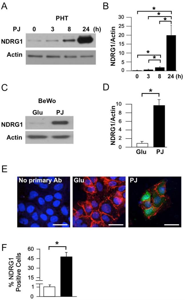
Figure 1. Effect of PJ on NDRG1 protein expression in primary human trophoblasts (PHTs) and BeWo choriocarcinoma cells.
Cells were cultured in 20% O2 for 24 h in the presence of glucose (Glu) or pomegranate juice (PJ). (A) Representative Western blot of NDRG1 in PHTs treated with PJ for the times indicated. (B) Quantification of NDRG1 levels, as represented in A (n=5 PHTs, *P ≤ 0.05 by ANOVA). (C) Representative Western blot of NDRG1 in BeWo cells treated with Glu or PJ for 24 h. (D) Quantification of NDRG1 levels in BeWo cells (n=3 experiments, *P ≤ 1.5 by Student's t-test). (E) Immunofluorescence of NDRG1 in PHTs stained for plasma membrane E-cadherin (red), nuclear DNA (blue), and NDRG1 (green). Scale bars: 10 μm. (F) Quantification of immunofluorescence, as represented in E. Percent of cells in which NDRG1 was detectable were 0% for no primary antibody 1.1±2% and 47.2±8.0% for glucose and PJ treated cells, respectively. (Mean±standard deviation, n=4 PHTs, *P ≤ 0.05 by Student's t-test).

