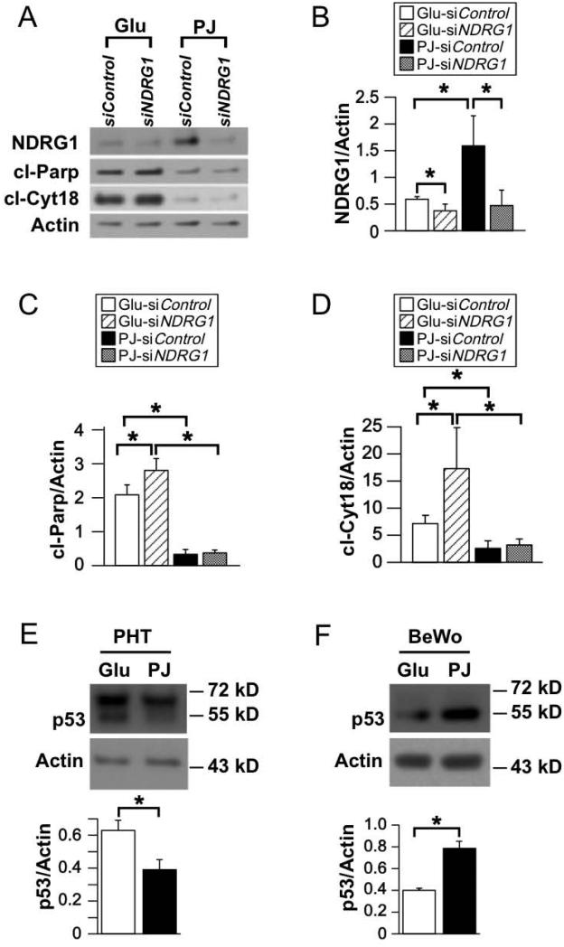
Figure 4. Effect of knockdown of NDRG1 on apoptosis in primary trophoblasts subjected to hypoxia in the presence of PJ or glucose.
Trophoblasts were cultured for 4 h in DMEM, transfected with siNDRG1 or scrambled siRNA control, treated with PJ or glucose for 8 h and then exposed to ≤1% O2 for 24 h while continuing PJ or glucose treatment. (A) Representative Western blots for NDRG1, cl-Parp, and cl-Cyt18 in transfected PHTs. (B-D) Quantification of NDRG1 (B), cl-Parp (C), and cl-Cyt18 (D), as represented in A. (n=4 PHTs, *P≤0.05 by Student’s t-test). (E, F) Representative Western blots (top) and quantification (bottom) for p53 in PHTs (E) and BeWos (F). (n=3 PHTs and n=6 experiments BeWos, *P ≤ 0.05 by Student’s t-test)

