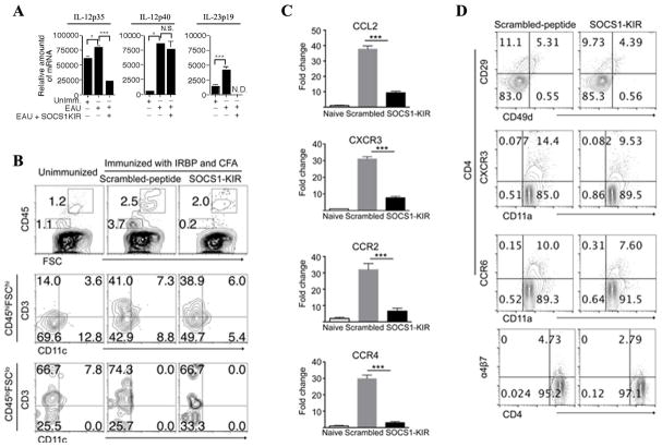
Figure 4. SOCS1-KIR suppresses trafficking of inflammatory cells into the retina.
(A) Retinae of mice treated with scrambled peptide or SOCS1-KIR were isolated on day 21 post-immunization, digested with collagenase and analyzed for the expression of IL-23 (IL-12p40, IL-23p19) or IL-12 (IL-12p35, IL-12p40) by qRT-PCR. (B) Flow cytometric analysis of inflammatory cells in the retina on day 21 after EAU induction. Numbers in quadrants indicate percentage of CD45+, CD3+ and/or CD11c+ cells in the retinae during EAU. Cells isolated from the spleen and LN of mice with EAU were re-stimulated in vitro with IRBP in medium containing SOCS1 KIR or scrambled peptides and analyzed for the expression integrins and chemokine receptors by qPCR (C) or FACS (D). Plots in (D) were gated on CD4+ T cells and numbers in quadrants indicate percent of CD4+ T cells expressing CD29, CD49d, CXCR3, CD11a, CCR6, or a4β7. Data are representative of three independent experiments.

