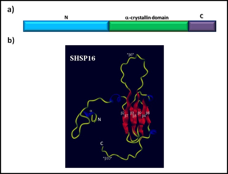
Fig. 1.
General structure of sHSPs. a Structural domains of the primary sequence represented by the amino terminal domain (N), the α-crystallin domain, and the carboxyl-terminal domain (C). b 3D model of the tertiary structure of SHSP16 protein from the protozoan parasite Trypanosoma cruzi. Ribbon model of the SHSP16 monomer generated with the SWISS-MODEL program. The crystallographic structure of the wheat HSP16.9 protein was used as a template to construct the model. The unstructured tails are shown in yellow, the α-helices in blue, and the β sheets in red. β sheets were numbered according to the corresponding sheets from the HSP16.9 structure; the sheets β6 and β10 (enclosed in quotes) are absent in SHSP16 of Trypanosoma cruzi. The N-and C-termini are labeled N and C, respectively (Color figure online)

