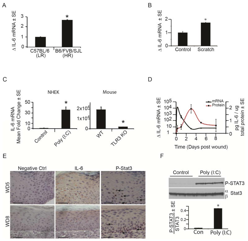
Figure 2. IL-6 and pSTAT3 are induced by tissue damage and double stranded RNA.
A) Mean fold change in IL-6 mRNA in healed scars at WD20-24 in HR vs. LR strains of mice as determined by qRT-PCR and normalized to housekeeping gene β-actin.
B) Mean fold change in IL-6 mRNA 24 hours post scratch assay in NHEK as determined by qRT-PCR and normalized to housekeeping gene, RPLP0.
C) Mean fold change in IL-6 mRNA after poly (I:C) addition (20μg/mL) to NHEK for 6 hours or in strain matched wt and TLR3 KO mice (n = 3) 6 hours after wounding as determined by qRT-PCR and normalized to housekeeping gene RPLP0 (NHEK) or β-actin (mouse).
D) Time course of IL-6 mRNA and protein expression throughout early stage wound healing in wt mice, as determined by qRT-PCR and ELISA, respectively. N = 3 mice/time point
E) IL-6 (middle) and P-STAT3 (right; arrows) immunohistochemistry of healing scars at WD5 (top) and WD8 (bottom) in wt mice. Scale bar = 50 μm.
F) P-STAT 3 levels in NHEKs after poly (I:C) (20ug/mL) for 24 hours compared to vehicle control as measured by western blot and normalized to STAT3.
*p < 0.05 by Student’s T-test or Single Factor ANOVA.

