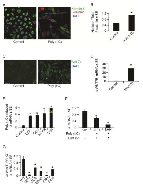
Figure 5. TLR3 activation induces hair follicle morphogenic program markers.
A) β-catenin immunofluorescence staining in NHEK after 72 hours of 24 hour treatment with poly (I:C) (20μg/mL) or control.
B) Quantitation of nuclear β-catenin to total levels of β-catenin in NHEK as in 5A.
C) WNT7b immunofluorescence staining (green) after 7 days of continuous poly (I:C)
(20mg/mL) or vehicle control treatment to NHEK. Scale bar = 50 μm; original magnification = 40X.
D) Mean fold change in Wnt7b mRNA after 6 days of continuous exposure to poly (I:C) (20mg/mL) determined by qRT-PCR and normalized to housekeeping gene, RPLP0.
E) Mean fold change in LEF1, GLI1, SHH and EDAR mRNA after poly (I:C) treatment by qRT-PCR as in 5A.
F) Mean fold change in LEF1 and SHH mRNA with TLR3-specific inhibitor or control in the presence of poly (I:C) in NHEK as determined by qRT-PCR as in 5A.
G) Mean fold change in Lef1, Edar, Gli2, Shh and β-catenin mRNA in TLR3 KO mouse wounds compared to strain-matched control mice as determined by qRT-PCR.
*p < 0.05 by Student’s T-test or Single Factor ANOVA.

