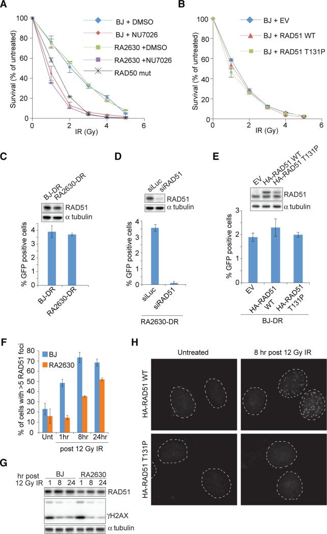
Figure 2.
Assessment of IR survival and homology directed repair (HDR) of cells expressing RAD51 T131P. (A) IR sensitivity of cells pretreated with NU7026 or DMSO. (B) IR sensitivity of BJ fibroblasts expressing RAD51 wild type (WT) or T131P. (C-E) GFP HDR assay in indicated cells with a stably integrated DR-GFP reporter. BJ-DR, and RA2630-DR (C), RA2630-DR cells depleted of RAD51 (D), and BJ-DR cells stably expressing HA-FLAG tagged WT or T131P RAD51 (E) were assessed. Error bars: s.d. (n=3). (F) Blinded quantification of cells with more than five RAD51 foci after IR. Error bars: s.d. (n=2). (G) RAD51 and γH2AX expression levels after IR. (H) IR-induced foci formation of HA-FLAG tagged RAD51 proteins. Dashed lines mark outline of nuclei. Also see Figure S2.

