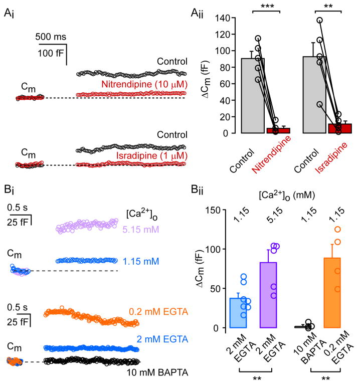
Figure 2. L-type Ca2+ current evokes exocytosis in AII amacrine cells.
Ai: Capacitance changes in AII-amacrine cells are inhibited by nitrendipine (10 μM) and isradepine (1 μM). Typical depolarization-evoked (500 ms; from −80 to −10 mV) exocytosis (ΔCm) from an AII-amacrine cell under control conditions (black) and after bath application of nitrendipine or isradepine (red). Aii: Bar graph summarizing ΔCm under control conditions and after drug application. Individual cells are represented by open circles. Experiments were performed at 31°C. Statistical significance (paired t-test) is denoted by asterisks. Bi: Top: Average ΔCm traces evoked by depolarization (100 ms pulse from −80 to −10 mV) from AII-AC under different external Ca2+ concentrations (1.15mM: n=6; 5.15mM: n=5). Bottom: Average ΔCm traces evoked by depolarization (100 ms pulse from −80 to −10 mV) from AII-AC for various internal Ca2+ buffer concentrations (10 mM BAPTA: n=4; 2 mM EGTA: n=6; 0.2 mM EGTA: n=5). Bii: Summary plot of ΔCm from individual AII-AC in various external calcium concentrations and internal calcium-buffers. Experiments were conducted at room temperature. Bar graphs denote mean ± SEM. Statistical significance (unpaired-t test) is denoted by asterisks.

