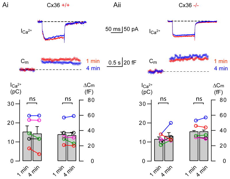
Figure 3. ΔCm in wildtype and Cx36(−/−) knockout mice.
Ai & Aii: Sample recordings of depolarization evoked Ca2+ currents (ICa) and resultant changes in membrane capacitance (Cm) from AII amacrine cells of a wild type (Cx36 +/+, Ai) and connexin 36 knockout (Cx36 −/−, Aii) mouse. Top traces show the Ca2+ current elicited by a 100 ms depolarizing pulse from −80 to −10 mV at 1 min (red) and 4 min (blue) after break-in. The ΔCm jumps are shown below. The bottom panels show summary graphs of ICa2+ charge and corresponding ΔCm from several cells at each condition. Bar graphs denote mean ± SEM.

