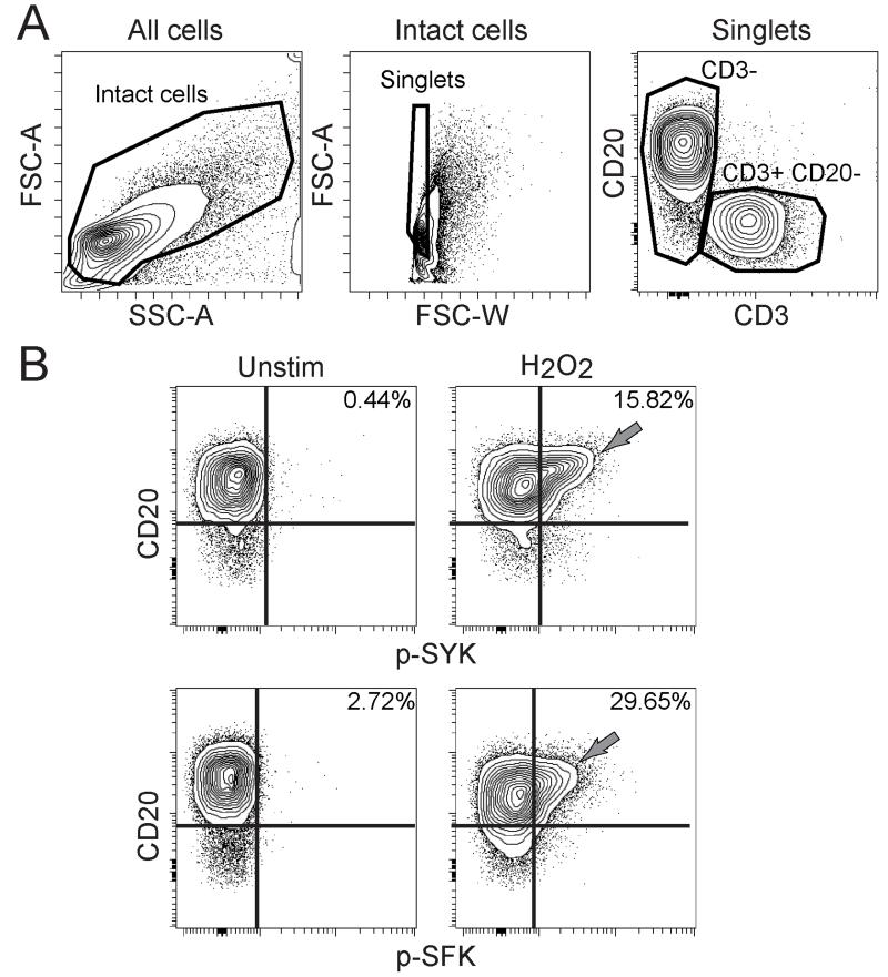
Figure 1. CD20hi B cells in human tonsil were sensitive to H2O2.
(A) Contour plots show gating for CD3− cells and CD3+ CD20− T cells in human tonsil. (B) Contour plots show phosphorylated SYK (p-SYK) and p-SFK in CD3− tonsillar B cells left unstimulated or stimulated by 3.3 mM H2O2 for 2 minutes. Sensitivity to H2O2 in a CD20hi B cell population is indicated (gray arrows). Plots are representative of three tonsils.

