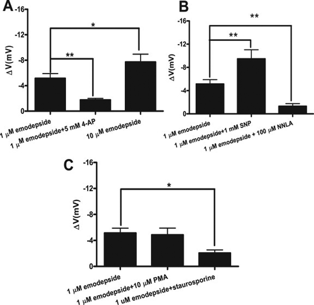
Fig. 4.

Effect of 1 µM emodepside on membrane potential. A: Comparison was made between membrane potential before and during emodepside application. 1 µM emodepside caused a significant membrane hyperpolarization (p < 0.001, n = 10, paired t-test) which was reduced in the presence of 5 mM 4-aminopyridine (p < 0.01, n = 8, unpaired t-test). 10 µM emodepside caused an increased hyperpolarization in comparison to 1 µM emodepside (p < 0.05, unpaired t-test). B Bar chart (mean ± s.e) of NO influence on emodepside-induced hyperpolarization. In the presence of 1 mM SNP, 1 µM emodepside caused an increased hyperpolarization (p < 0.01, n = 4, paired t-test). 100 µM NNLA decreased the hyperpolarization caused by 1 µM emodepside (p < 0.01, unpaired t-test). C: Bar chart (mean ± s.e) of effect of protein kinase modulators on emodepside-induced hyperpolarization. 10 µM PMA had no significant effects on the hyperpolarization caused by 1 µM emodepside. However, 1 µM staurosporine decreased the hyperpolarization caused by 1 µM emodepside (p < 0.05, unpaired t-test). (Martin et al., 2012)
