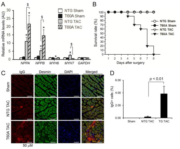
Figure 5. Effects of CR-PsmI on cardiac responses to sTAC.
A, Changes in the mRNA expression of the indicated genes at 6 days after surgery. GAPDH was probed as a house keeping gene for RNA quantification control. *p<0.05 vs. NTG sham; $p<0.05; n=4 mice/group. B, Kaplan-Meier survival curve. n=10 mice/group; p < 0.01, log-rank test. C & D, Prevalence of cardiomyocyte necrosis. Cryosections from LV myocardium collected 6 days after sTAC were immuno-stained for mouse IgG (red) using a Fluor-568 conjugated anti-mouse IgG antibody and for desmin using rabbit primary antibodies for desmin and Fluor-488 conjugated anti-rabbit IgG secondary antibodies. Desmin- and mouse IgG positive (IgG+) cardiomyocytes are considered necrotic. Nuclear DNA was stained blue with DAPI. Representative images (C) and pooled data from 3 mice per group (D) are shown.

