
Figure 2.
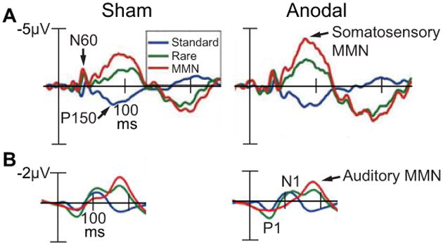
A, Event-related potentials (ERPs) recorded during a vibratory somatosensory discrimination task following 25 min of tDCS over the right cerebellar hemisphere. ERPs elicited from vibratory standard stimuli (blue), rare stimuli (green), and the difference between standard and rare stimuli (i.e., the somatosensory mismatch negativity or MMN, red) shown at the left centrolateral electrode (C3) across sham and anodal conditions. Arrows show the N60, P150, and somatosensory MMN. B, ERPs recorded during an auditory discrimination task following 25 min of tDCS over the right cerebellar hemisphere. ERPs elicited from auditory standard stimuli (blue), rare stimuli (green), and the difference between standard and rare stimuli (i.e., the auditory mismatch negativity or MMN, red) shown at the left centrolateral electrode (C3) across sham and anodal conditions. Arrows show the P1 and N1 elicited from the standard auditory stimulus (blue), and the auditory MMN. Adapted from Chen et al (2014) with permission from The Physiological Society.