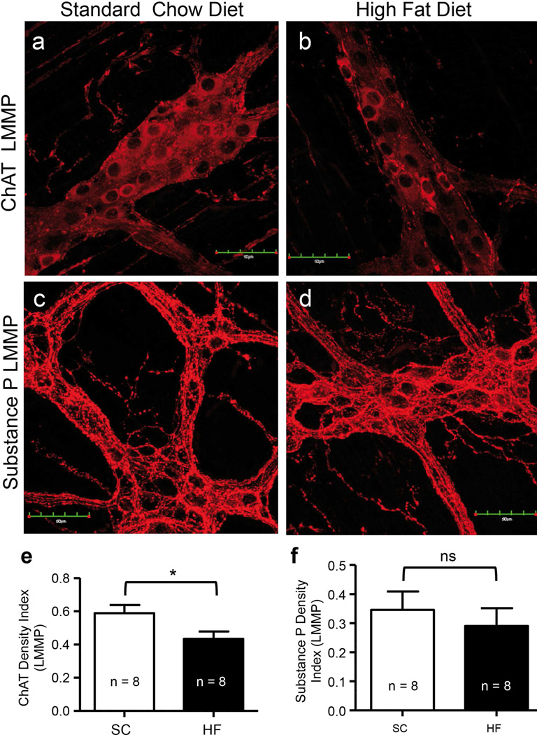
Fig. 5.
Myenteric ganglia excitatory neurons were variably affected by the ingestion of a HF diet for 20 weeks. Duodenal LMMP preparations from 20-week SC and HF diet mice showing ChAT-IR (a, b) and substance P-IR (c, d) in the myenteric plexus of mouse duodenum. ChAT density indices were reduced by a HF diet (e; P = 0.039; n = 8, unpaired T-test), but substance P density indices were not different between 20-week SC and HF diet mice (f; P = P = 0.546; n = 8, unpaired T-test).

