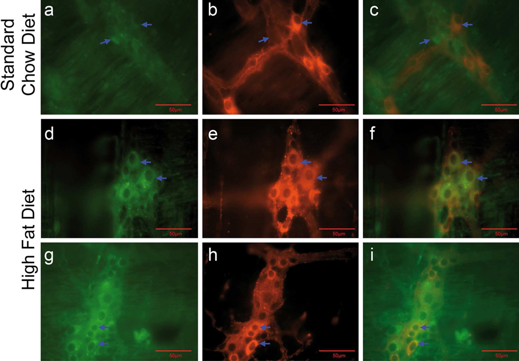
Fig. 7.
A subpopulation of duodenal myenteric neurons of mice fed a HF diet for 20 weeks exhibited activated apoptotic pathways. LMMP preparations were used to demonstrate co-localization of cleaved caspase-3-IR with nNOS-IR (a–f) and ChAT-IR (g–i) neurons in the myenteric ganglia. Compared to mice fed a SC diet for 20 weeks (ac; caspase-3 + nNOS), those fed a HF diet showed caspase-3-IR in nNOS-IR nerve cell bodies (d-f; caspase-3 + nNOs) and in ChAT-IR nerve cell bodies (g-I; caspase-3 + ChAT) indicating an activation of apoptosis in nNOS and ChAT neurons. Arrows of a-c indicate a lack of co-localization of caspase-3 and nNOS-IR. Arrows in d-f and g-I show co-localization of these markers (d–f), as well as co-localization of caspase-3 and ChAT (g–i). J, Caspase-3-IR nerve cell bodies per ganglion area were computed before staining the preparations to localize caspase-3-IR with nNOS and ChAT. Summary data show an increased number of myenteric caspase-3-IR nerve cell bodies per ganglion area in mice fed a HF diet for 20 weeks (P= 0.0017, n = 5, unpaired t-test) compared to mice fed a SC diet.

