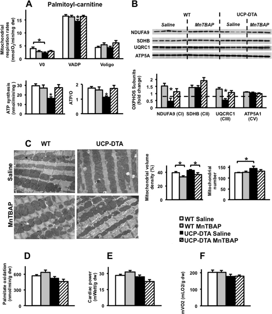
Figure 4. Impact of MnTBAP on mitochondrial function and myocardial substrate metabolism in 24 week-old UCP-DTA mice.
A. Oxygen consumption, ATP synthesis, and ATP/O ratios measured using saponin-permeabilized LV fibers with palmitoyl-carnitine and malate as substrates (n=6–8). B. Abundance and densitometric analysis of selected OxPhos subunits in the whole heart homogenates as measured by western blotting (n=4–6). C. Electron microscopy images (1:10,000) and quantification of mitochondrial number and volume density in left ventricular sections of 24 week-old UCP-DTA mouse hearts (n=4). D–F. Palmitate oxidation, cardiac power and mVO2 measured ex vivo in isolated working hearts (n=6–8). *p<0.05 vs. all other or indicated groups.
Dashed line or  Saline-treated control wildtype (WT) mice,
  MnTBAP-treated WT mice,
  UCP-DTA treated with saline,
  UCP-DTA treated with MnTBAP. Dw=dry weight.

