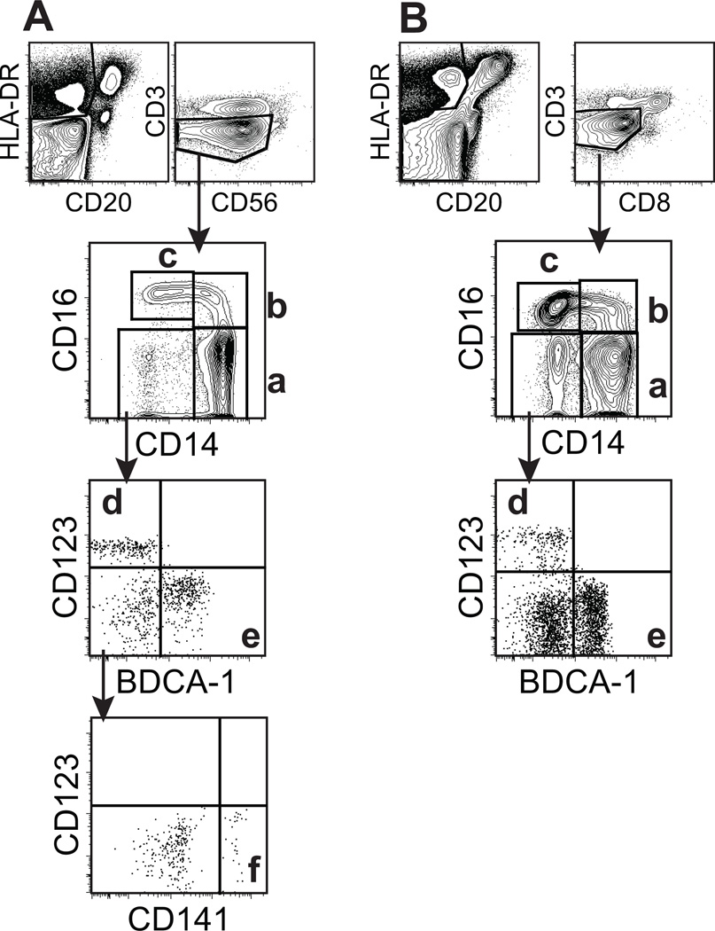
Figure 1. Phenotying of blood monocyte and DC subsets in humans (A) and rhesus macaques (B).
EDTA-treated blood samples were stained with antibodies shown in Table 1 and analyzed by 11-color flow cytometry. (A) In HLA-DR+CD3−CD20−CD56− populations, human monocyte and DC subsets were gated and divided into 4 populations by CD14 and CD16 expression as follows; (a) CD14+CD16− monocytes, (b) CD14+CD16+ monocytes, (c) CD14−CD16+ monocytes, and a CD14−CD16− population that was further divided into (d) CD123+ pDC and (e) BDCA-1+ mDC. In addition, a CD141+ mDC (f) was identified. (B) To analyze rhesus monocyte and DC subsets, HLA-DR+CD3−CD20−CD8−cell populations were similarly gated and further divided as described in panel A with the exception that antibody to human CD141 (BDCA-3) did not cross-react to, or detect this marker on rhesus macaque cells. The populations of cells identified included; (a) CD14+CD16− monocytes, (b) CD14+CD16+ monocytes, (c) CD14−CD16+ monocytes, (d) CD123+ pDC, and (e) BDCA-1+ mDC.

