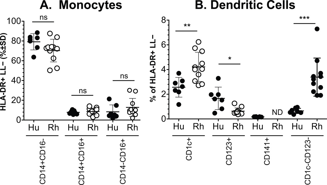
Figure 2. Comparison in the proportion of blood monocyte and DC subset populations between humans and rhesus macaques.
Blood from 7 humans and 11 rhesus macaques were gated and analyzed as described in the materials and methods and in Figure 1. The proportions of monocytes (A) and DC (B) subsets were calculated from the total number of HLA-DR+ lymphocyte lineage (LL) negative cells. Comparisons between cell subsets in human and rhesus macaque blood were calculated for statistically significant differences by the Mann Whitney rank test. Not detected, ND; P<0.05,*; P<0.01,**; P < 0.001,***.

