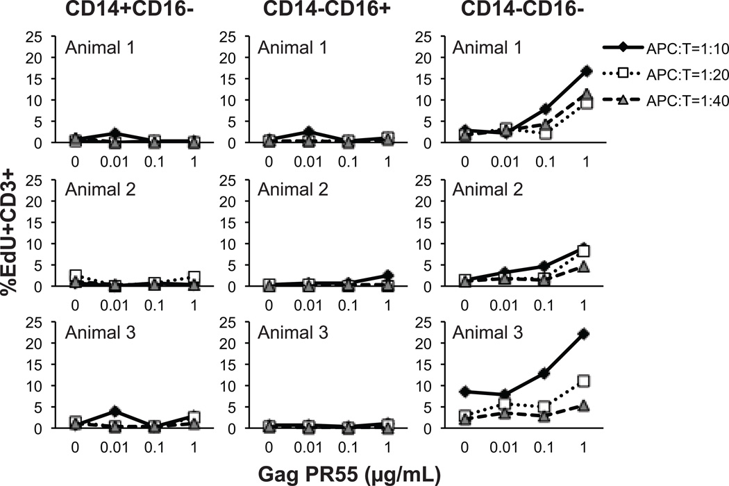
Figure 4. Cell proliferation induced by antigen presentation on rhesus monocyte and DC subsets.
CD14+CD16− classical monocytes, CD14−CD16+ non-classical monocytes, and the CD14−CD16− fraction that includes DC subsets were sorted by flow cytometry of blood samples obtained from SIV-infected and ART-treated rhesus macaques. The subset populations were then pulsed with Gag pr55 protein at indicated concentrations for 2 hr. The antigen-pulsed effector cells were added to wells at numbers ranging from 2500 - 10000 per well and incubated with 1×10⁵ autologous CD3+ T cells per well for 4.5 days resulting in antigen presenting cell to T cell (APC:T) ratios of 1:40-1:10. The thymidine analogue, EdU, was added during the last 18 hr of incubation and EdU incorporation in CD3+ T cells was detected by immunostaining and flow cytometry. Data from three animals are shown.

