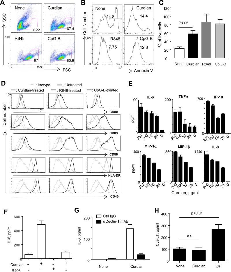
FIGURE 2.
hDectin-1 ligation by β-glucan activates pDCs via Syk. pDCs from blood were cultured overnight in the presence of none, curdlan, R848, or CpG-B. (A) FSC and SSC scatter gram of pDCs. (B) Annexin V staining of cells gated in (A). (C) Summary of three independent experiments of A and B. Error bars represent SD. (D) Expression levels of CD80, CD83, CD86, HLA-DR, and CD40 were measured. (E) Cytokine and chemokine levels. (F) pDCs were treated with a Syk inhibitor, R406, for 1h and then cultured overnight with 100 μg/ml curdlan. The amount of IL-6 in the culture supernatants was assessed. (G) pDCs were treated with 20 μg/ml anti-hDectin-1 antibody and then incubated overnight with 100 μg/ml curdlan. The amount of IL-6 in the culture supernatants was measured. In (E-G), error bars represent SD of triplicate assays. Two independent experiments showed similar results. (H) The amount of cysteinyl leukotriene (Cys-LT) secreted from pDCs stimulated with curdlan or house dust mite Dermatophagoides farina (Df) extract. FACS-sorted pDCs from two healthy donors were tested in duplicate assays (Mean ± SD).

