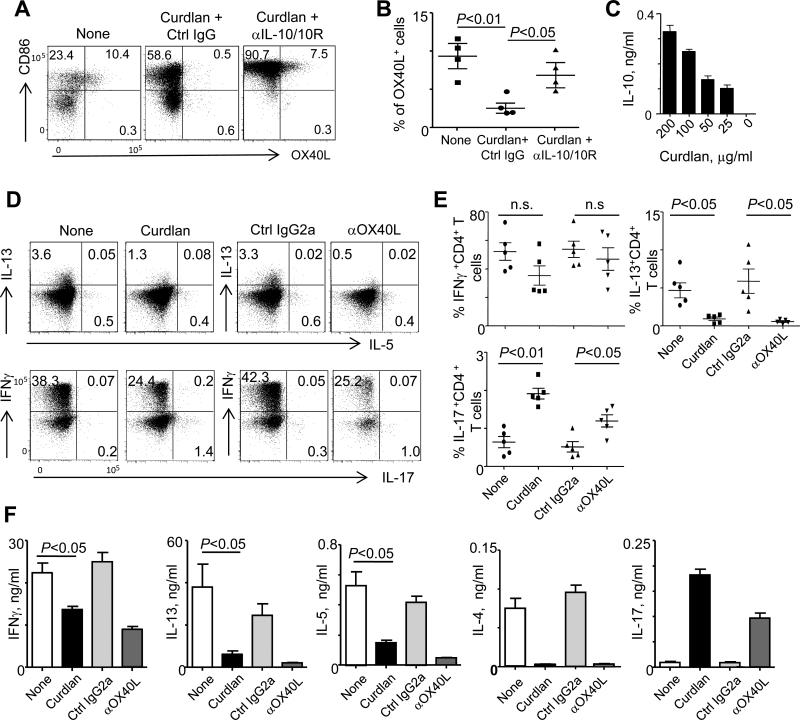
FIGURE 4.
hDectin-1-activated mDCs decrease Th2 induction and Th2-type cytokine secretion by CD4+ T cells. (A) Blood mDCs were cultured overnight in medium alone or either in the presence of curdlan and control IgG or curdlan and anti-IL-10/IL-10R. Cells were stained with anti-CD86 and anti-OX40L antibodies. (B) Summarized data of (A) from four independent experiments using cells from different donors. (C) Levels of IL-10 secreted from mDCs cultured overnight with the indicated amounts of curdlan. Error bars indicate SD of triplicate assays. Two independent experiments showed similar results. (D) Allogeneic naïve CD4+ T cells were co-cultured for 7 days with untreated pDCs and curdlan-treated pDCs (left panels) in the presence of control IgG or anti-OX40L antibody. T cells were stained for intracellular IL-13/IL-5 (upper panels) as well as IFNγ/IL-17 (lower panels) expression during stimulation with PMA/ionomycin. (E) Summarized data of experiment (D) performed with cells from different donors. (F) Cytokines secreted from T cells (E) during 48 h restimulation with PMA/ionomycin. Error bars indicate SD of triplicate assays. Two independent experiments showed similar results. ANOVA for (B) and (E) and t-test for (F) were used for testing statistical significance of the data.

