
Figure 3.
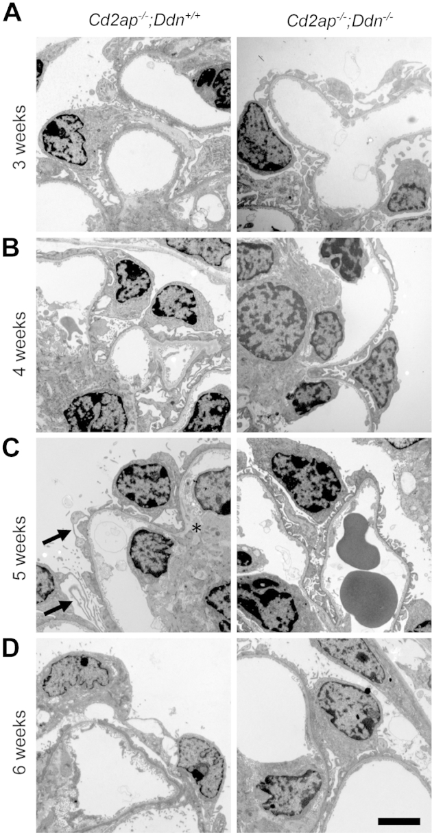
Dendrin ablation ameliorates podocyte injury in Cd2ap−/− mice. A: At 3 weeks, Cd2ap−/−;Ddn+/+ mice show widespread foot process broadening (FPB), segmental foot process effacement (FPE), microvillous transformation of the podocyte cell membrane, and focal vacuolization of podocytes. By contrast, Cd2ap−/−;Ddn−/− mice show only mild FPB at 3 weeks. B: Although Cd2ap−/−;Ddn+/+ mice show widespread FPE at 4 weeks, podocyte foot processes are still mostly preserved with very focal FPE and FPB in Cd2ap−/−;Ddn−/− mice. C: At 5 weeks, podocytes of Cd2ap−/−;Ddn+/+ mice show diffuse FPE, widespread microvillous transformation of their cell membrane, and features of hypertrophy, characterized by thin and long processes (arrows). In addition, an expanded mesangial matrix infiltrates the area between the endothelium and the GBM (asterisk). The glomerular endothelium shows mild loss of fenestrations. By contrast, there is only segmental FPE and extensive FPB in 5-week-old Cd2ap−/−;Ddn−/− animals. D: At 6 weeks, both Cd2ap−/−;Ddn+/+ and Cd2ap−/−;Ddn−/− mice show widespread FPE; however, in the Cd2ap−/−;Ddn+/+ mice, areas of denuded GBM are frequently identified. Scale bar = 2 μm.